Dental X-Rays
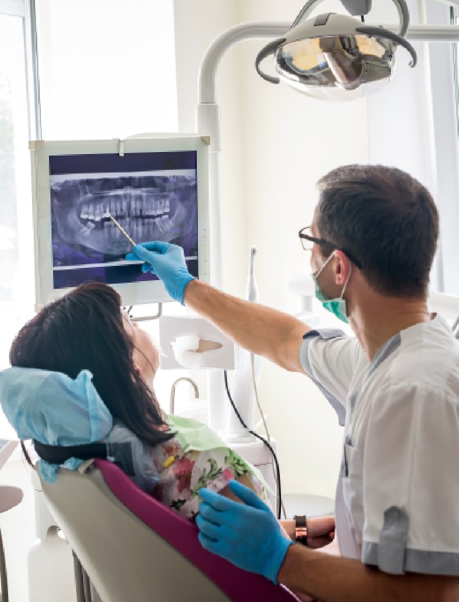
Dental x-rays are an efficient and effective tool used in dentistry to evaluate oral health. They are used to identify a number of dental issues related to teeth, gums, and/or bone. Breakthroughs in dental technology, like low radiation digital x-rays, continue to make the procedures we perform safer and more precise in evaluating our patients’ dental health. At Rockefeller Cosmetic Dentistry, comfort and expertise are our priorities, which is why we’ve chosen to utilize low radiation digital radiography methods.
Types Of Dental X-Rays
There are two main types of dental x-rays utilized for imaging: intraoral and extraoral. The most common are intraoral x-rays, which capture images of the inside of the mouth. Intraoral imaging helps to determine the health of the roots, cavities in the teeth, and evaluate the health of the bone and jaw. There are several types of intraoral x-rays, each capture different angles of the mouth:
Bitewing X-rays
Bitewing x-rays capture images of the upper and lower teeth in one distinct area of the mouth. It shows the crown of the tooth, to the level of supporting bone. This type of image is used to detect any decay in between the teeth or any deterioration of the bone.
Periapical X-rays
Periapical x-rays show the entire mouth, from the crown to where the root attaches into the jaw bone. These images are used to determine if there is any abnormal changes to the tooth root or surrounding bone.
Occlusal X-rays
Occlusal x-rays are a diagnostic tool used to track the development of the entire arch of the upper or lower mouth.
Extraoral imaging focuses on the bone and tissues of the skull and jaw. These images allow our specialists to view full images of the patient’s skull. Types of extraoral imaging include:
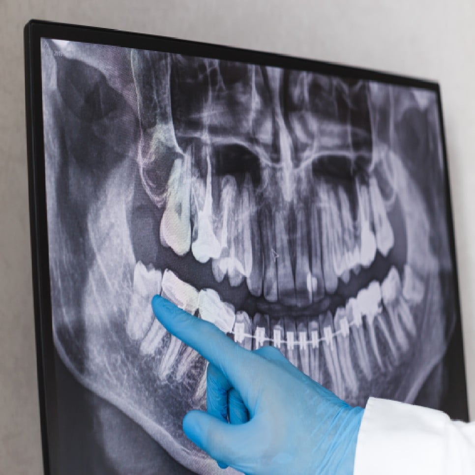
Panoramic X-rays
This type of imaging captures the entire mouth region in one single image. Panoramic x-rays are useful in detecting fully emerged, newly emerged, impacted teeth, and help diagnose tumors.
Dental Computed
Tomography (CT scanning)
This is a 3D image of the inside of the mouth to identify issues in the bone structure, such as tumors or fractures. They also help evaluate bone strength for the placement of dental implants and difficult extractions.
Cone Beam CT (CBCT)
Cone Beam CT scans provide high quality 3D images of the soft tissue, nerves, and bone. This type of imaging acts as a guide for dental implant placement, as well as can detect any complications in the gums, tooth root, and jaws.
Dental x-rays are typically performed during the initial exam. Depending on the issues or severity of the case, different types of images will be ordered in order to ensure a proper diagnosis. Combining both types of imaging, intraoral and extraoral, provides specialists with a comprehensive report on the current state of the patient’s mouth.
Digital Dental X-Ray Benefits Include:
[faq id=”136″]
FAQ
[faq id=”105″]
X-Ray Uses In Dentistry
Dental x-rays are often taken routinely in order to detect dental issues in their early stages, even prior to experiencing any symptoms. These images allow our specialists to detect:
- Any areas of tooth decay.
- Bone loss caused by periodontal disease.
- Changes in the root canal.
- Abscesses, which are infections at the root of the tooth or between the tooth and gum.
- Dental injuries or damage to the that supports the teeth.
Dental x-ray imaging is also used for treatment planning for cavities, root canal therapy, tooth extraction, and for the placement of dental implants.
The potential physical and environmental impacts of traditional film x-rays have long been the source of debate in every area of the health field. Unlike traditional film x-rays, our digital method reduces radiation exposure by up to 90%, making digital radiography the fastest, safest, and most precise type of imaging available. Due to the very minimal levels of radiation being exposed, the risk of potential harmful effects are very small. Dental x-ray techniques are designed to limit the body’s exposure to radiation and several precautionary methods are taken to ensure your safety and comfort. When performed with the proper precautionary techniques, there is very little cause for concern.